
Ultrasound time-harmonic elastography for the assessment of glomerulonephritis
Markus Großmann, Ingolf Sack, Stephan Rodrigo Marticorena Garcia
The kidney function is of vital importance for the human metabolism, blood circulation and endocrine system. Glomerulonephritides comprises a group of immunologic renal diseases that can result in structural damage and functional failure due to inflammatory processes in the kidney. Unfortunately, currently available noninvasive tests are barely capable to detect early stage kidney disease and invasive renal biopsies are unsuitable for screening purposes. Since chronic kidney damage is irreversible there is a notable interest for noninvasive diagnostic tools to identify early stage candidates and might open new perspectives for treating early disease stages.
The objective of this project is to adapt the ultrasound time-harmonic elastography (THE) for native kidneys to acquire full field-of-view maps of renal stiffness and to apply the THE as a noninvasive medical imaging for the detection of structural changes in glomerulonephritis.
In our study we enroll healthy volunteers and patients with biopsy-proven glomerulonephritis and stage 1 to 4 chronic kidney disease. We obtain full field-of-view maps of renal shear wave speed (SWS) to assess renal tissue stiffness. SWS will be analyzed separately in kidney substructures such as renal parenchyma, cortex and medulla to be correlated with clinical data, conventional ultrasound measurements and resistive indices (RI).
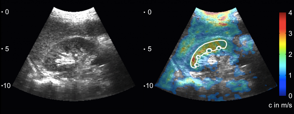
Figure: b-mode and elastogramm of a native kidney